Normalize the radiograph
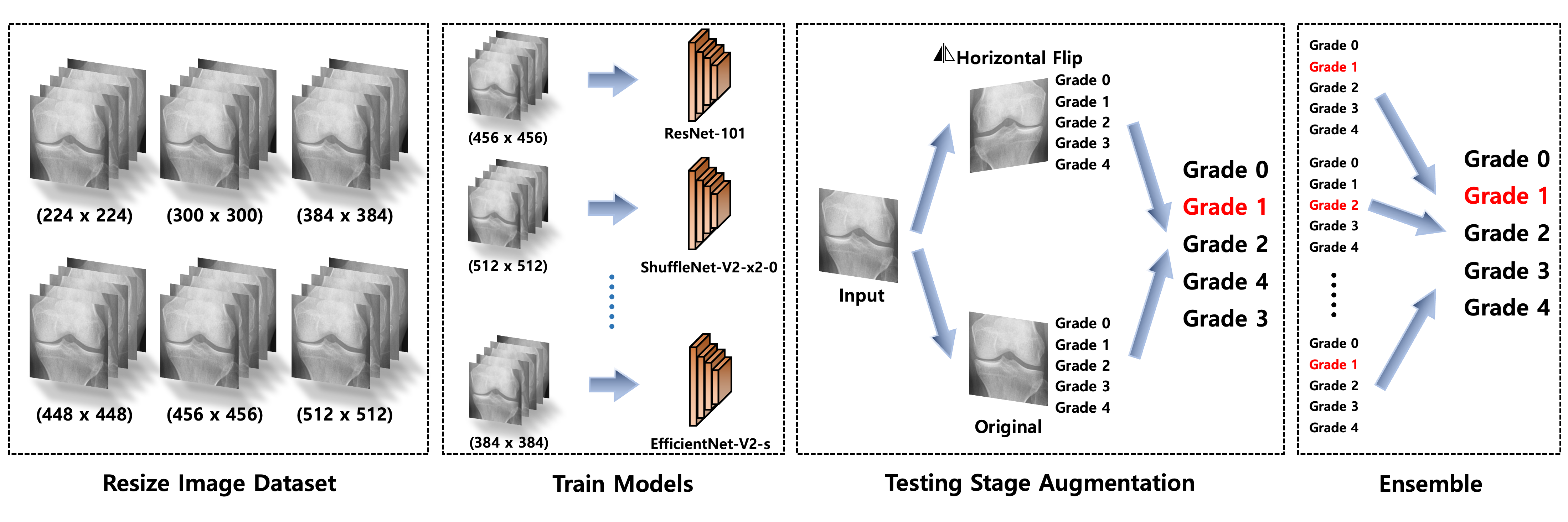
Each knee X-ray is prepared and resized into several model-specific resolutions.
 ORTHOGRADE AI
OrthoGrade AI combines eight complementary deep-learning networks to estimate Kellgren–Lawrence severity from knee radiographs—then reveals the joint regions that influenced the decision.
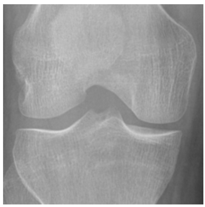
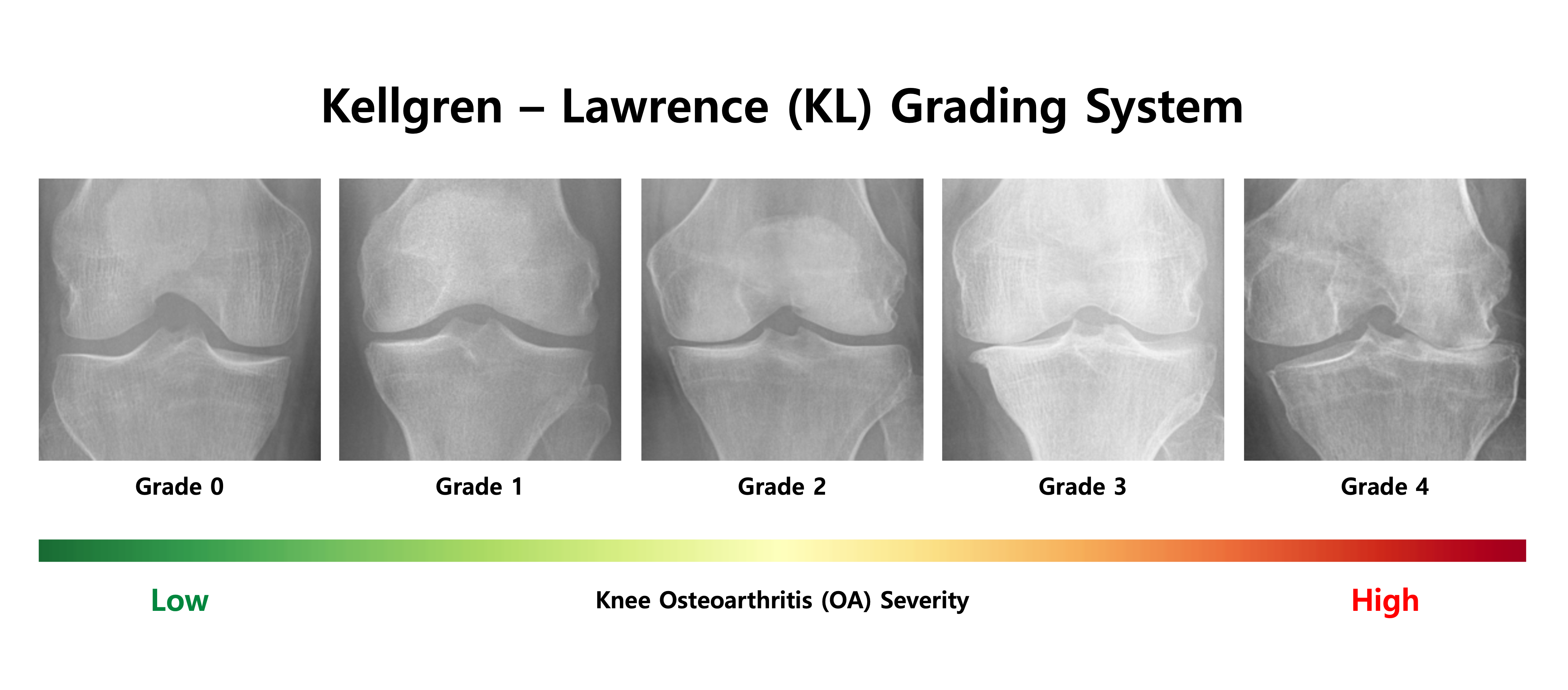
The Kellgren–Lawrence scale progresses from a radiographically normal joint to severe osteoarthritis. Select a grade to inspect the characteristic pattern and watch the dataset spectrum respond.
Normal-appearing joint space without definite osteophytes or structural narrowing.
Instead of trusting a single neural network, the system trains a portfolio of architectures at image sizes that suit each model. During inference, original and horizontally flipped views are combined through mixed hard-and-soft voting.
Each knee X-ray is prepared and resized into several model-specific resolutions.
Eight pretrained model families learn complementary texture, shape and joint-space cues.
The original image and a horizontal flip are evaluated to make predictions more stable.
Hard class votes and soft probabilities are fused into the final grade estimate.
Grad-CAM highlights the image regions contributing most strongly to the prediction.
Different network families receive images at resolutions selected for their own representational strengths, rather than forcing every model to use one common size.
Grad-CAM heatmaps provide a visual check that the network is attending to clinically meaningful regions—especially the tibiofemoral joint space and areas associated with osteophyte formation.
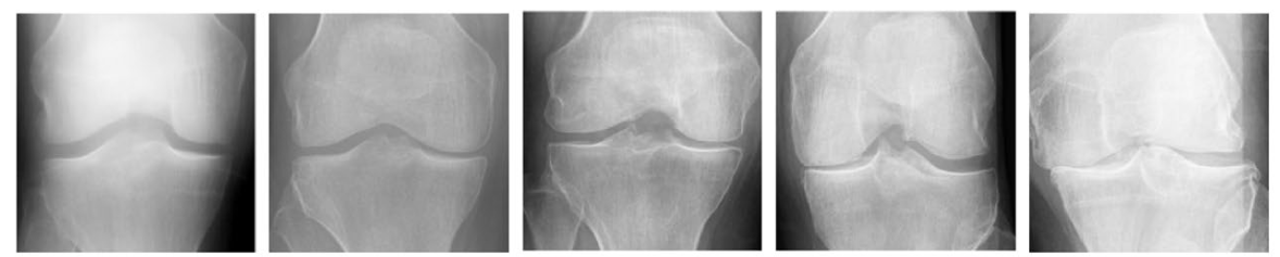
The gap between femur and tibia becomes progressively reduced as cartilage loss advances.
Marginal bony growths around the joint are a defining radiographic cue in KL grading.
Increased density and structural remodeling may appear with more advanced disease.
Severe grades can show marked contour change and loss of normal joint architecture.
The ensemble was evaluated on a held-out test set after stratified five-fold cross-validation. Results below reflect the reported experimental study and are not a claim of clinical deployment performance.
Balance between precision and recall across five ordered classes.
Posterior–anterior fixed-flexion knee radiographs across KL grades 0–4.
Held apart from cross-validation and model training.
The 6,604 training-and-validation images are divided into five class-balanced folds. Four folds train the models while one validates them, rotating until every fold has served as validation.
For a demonstration environment, the workflow can sit beside a digital radiography system: receive a knee image, estimate the KL grade, expose the attention map, and produce a standardized summary for clinician review.
This website demonstrates a published experimental AI workflow. It is not a medical device and must not be used as the sole basis for diagnosis, treatment or patient management.
Objective grading. Visible evidence. A presentation built for clinical, laboratory and instrument stakeholders.
Powered with ❤️ by Solicitous ®