Solicitous logo Solicitous®Clinical AI Systems
Open Workstation
Multimodal Oncologic Imaging

OncoFusion AI

3D PET/CT Head & Neck Tumor Segmentation

A presentation-ready research workflow that combines anatomical CT structure with metabolic PET activity to delineate gross tumor volume across a standardized 3D scan.

Powered with ❤️ by Solicitous ®
3Dvolumetric analysis
PET + CTmultimodal input
144³standardized ROI
Fusion Review / Case HN-042 AI READY
PET and CT head and neck tumor views
Active modalityPET/CT Fusion
Voxel grid1 mm isotropic
Review stateSegmentation visible
Metabolic hotspot
GTV contour
ANATOMY-AWAREMETABOLICALLY INFORMED3D VOLUMETRICEXPERT REVIEWABLE
The clinical logic

What is actually happening?

The model is not simply highlighting a bright spot. It aligns two complementary imaging signals, normalizes them, focuses on the head-and-neck region, and predicts a 3D gross-tumor-volume mask.

CT

CT provides structure

Computed tomography contributes anatomy: bone, airway, soft tissue boundaries and spatial context.

PET

PET provides activity

Positron emission tomography shows radiotracer uptake, helping reveal metabolically active tumor tissue.

F

Fusion provides context

The network learns where uptake sits relative to anatomy, reducing reliance on either modality alone.

3D

The output is a volume

Instead of one image-level label, the output is a voxel-level tumor mask spanning the scan volume.

Interactive modality theatre

Normalization changes what the network sees

Switch between CT, raw PET and two PET normalization strategies. The animation preserves the original panels and overlays a scan cursor without cropping the medical image.
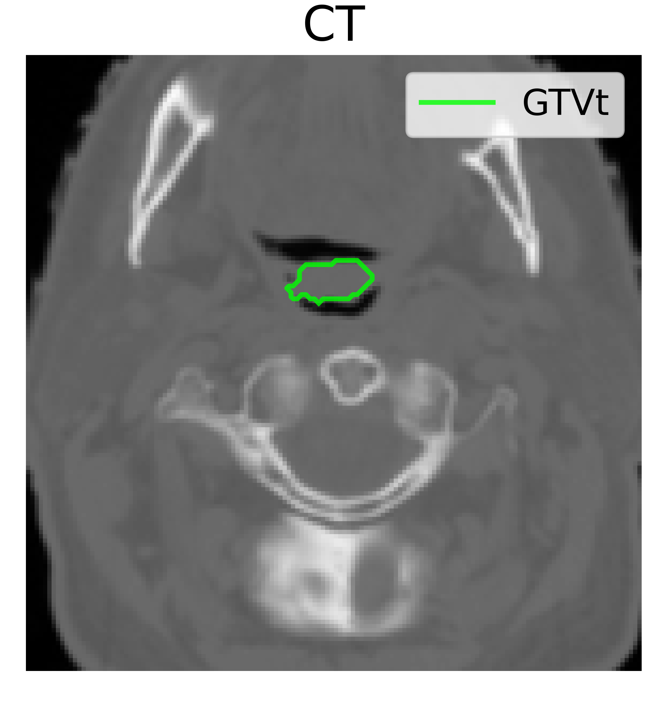
CT Anatomy
AXIAL 072 / 144
CT modality image
GTV
Anatomical reference for localization
End-to-end workflow

From scanner data to a reviewable tumor volume

STEP 01 / DATA INGEST

Load paired PET and CT studies

The workflow starts with co-registered imaging volumes. CT provides structural anatomy while PET contributes metabolic activity.

DICOM / NIfTIMultimodal3D
Smart section

PET normalization laboratory

Normalization can suppress distracting uptake and reshape intensity contrast. This matters because a segmentation model learns from the visual distribution it receives.
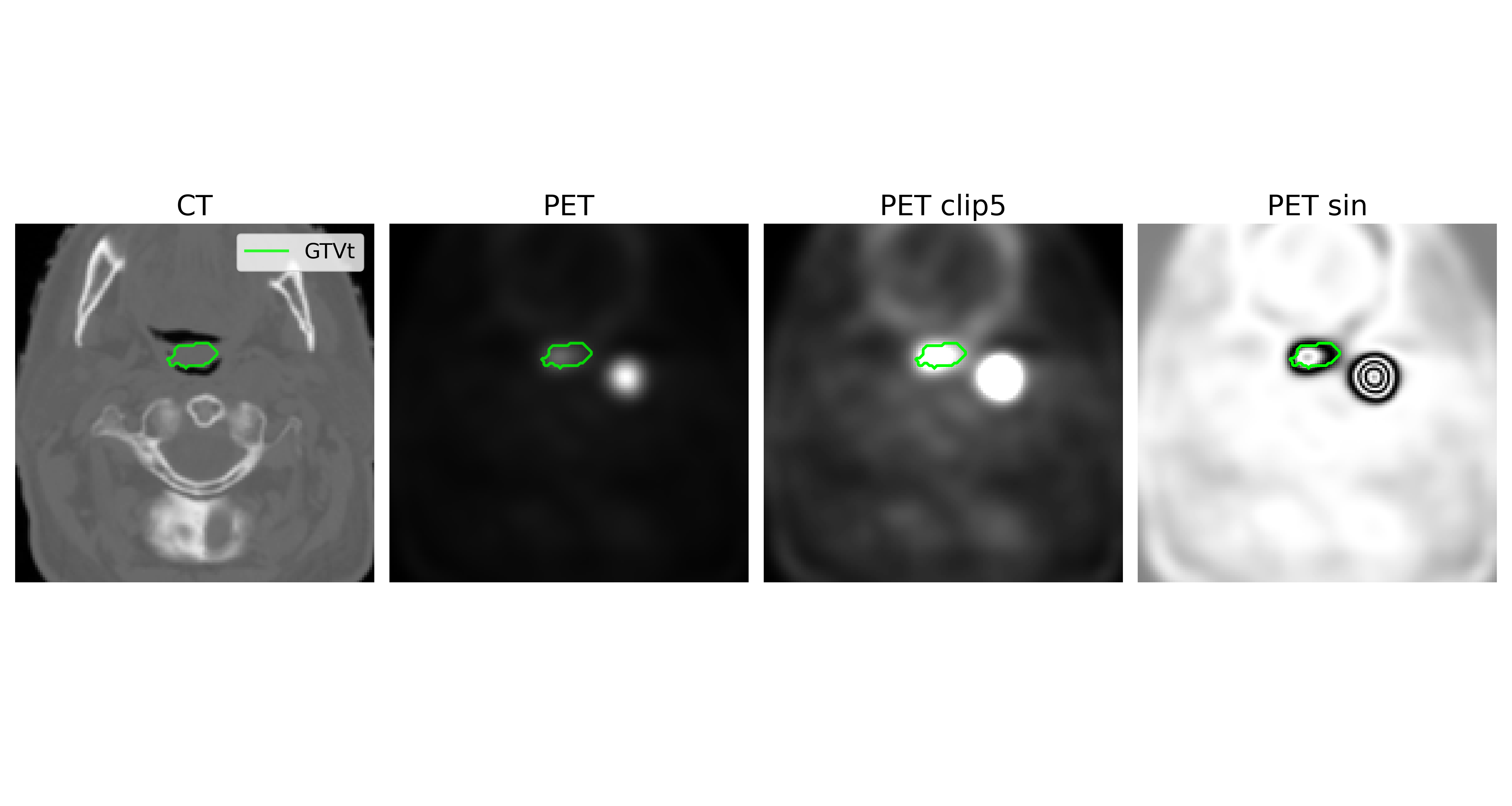
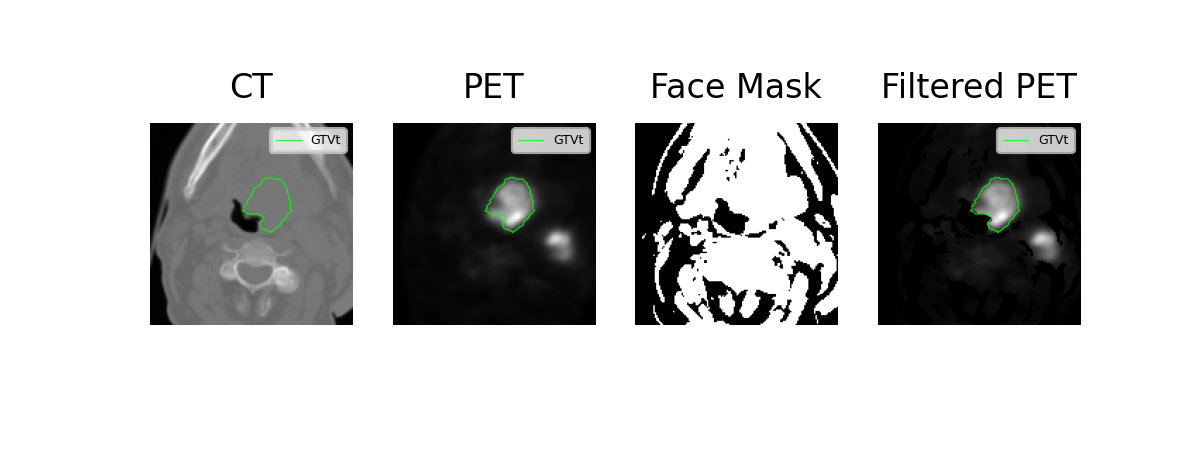
PET clipping workflow
Clipping strategyLimits extreme uptake values so the tumor contour receives stronger relative contrast.
Face mask filtered PET workflow
Face-mask filteringUses a head-and-neck mask to reduce irrelevant activity outside the anatomical region of interest.
Transformation simulatorPET Clip

Clipping compresses extreme hotspots while preserving a strong lesion-to-background contrast.

Visual evidence

Same patient, different preprocessing, different contour behavior

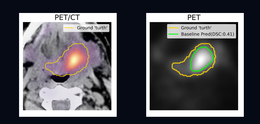
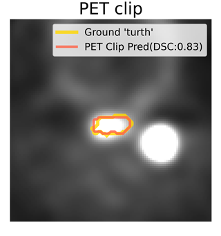
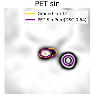
The project figures compare expert ground truth with predictions from baseline PET and transformed PET channels. Values shown are example-case Dice scores from the supplied figures, not a universal clinical performance claim.
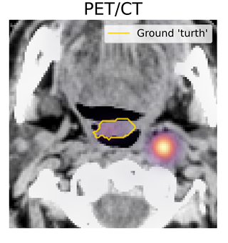
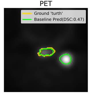
PET CT ground truth
PET/CT ReferenceExpert contour
Baseline PET prediction
Baseline PETDSC 0.47
PET clip prediction
PET ClipDSC 0.83
PET sin prediction
PET SinDSC 0.54
Smart interpretation

For this compact lesion, clipping improves contour agreement in the supplied example by compressing the competing high-intensity hotspot.

Model engine

3D segmentation core

The source workflow uses nnU-Net as the baseline framework for full-resolution 3D gross-tumor-volume segmentation and evaluates models through five-fold cross-validation.

CT
PET
+feature fusion
ENCODER
3D latent
representation
DECODER
GTV Maskvoxel-wise output
Why 3D?Tumors extend across slices; volumetric context helps maintain continuity.
Why multimodal?PET and CT answer different questions and become more useful when interpreted together.
Why cross-validation?Five-fold evaluation tests stability across multiple train/validation splits.
Beyond segmentation

Imaging features can support treatment-outcome research

The source project also contains a separate treatment-outcome prediction track. Segmentation can define the tumor region from which radiomic or learned features are extracted for downstream research such as progression-free-survival prediction.

Research support — not a diagnostic or prognostic device
1Segment tumor
2Extract features
3Build model
4Review risk signal
Instrument-connected vision

How this becomes a compelling local demo

01

Scanner ingestion

Load paired PET and CT studies from a local demonstration folder.

02

Automatic preprocessing

Resample, crop and normalize with visible progress and quality checks.

03

Interactive contour review

Inspect the mask over CT, PET and fusion views before accepting it.

04

Quantitative report

Summarize tumor volume, slice span, centroid and modality observations.

Solicitous
OncoFusion AI Analysis SummaryDemonstration report · Case HN-042
RESEARCH USE
Detected volume12.8 cm³Illustrative UI value
Slice coverage31 slicesAxial extent
Primary modalityPET/CTFused interpretation
Review statusPendingExpert confirmation required
Solicitous logo
AI-assisted multimodal oncology imaging

See anatomy. See activity.
Understand the volume.

Powered with ❤️ by Solicitous ®

Return to Top ↑