
Pixel-level localization
Semantic masks outline tumor sub-regions instead of only indicating that an abnormality exists.
A presentation-ready deep-learning workflow for converting multi-modal brain MRI volumes into clear, pixel-level tumor-region maps.
Turn complex MRI volumes into a visual narrative that is faster to review, easier to explain and more consistent across cases.
Semantic masks outline tumor sub-regions instead of only indicating that an abnormality exists.
3D volumes are decomposed into trainable slices across axial, coronal and sagittal views.
Original scan, reference mask and prediction can be reviewed side by side without hiding image boundaries.
Each sequence emphasizes different tissue characteristics. Together, they give the model richer context for separating tumor regions from normal anatomy.
Anatomical detail with fluid appearing relatively dark.
Water-rich tissue and edema appear with stronger signal intensity.
Contrast enhancement highlights regions with blood-brain barrier disruption.
Suppresses cerebrospinal fluid to improve visibility of edema and lesions.
The original four-sequence reference strip is kept completely visible with object-fit: contain; no scan edges are cropped.
A simple color system makes the final prediction easier to interpret during a presentation.
Non-tumorous pixels forming the contextual anatomy around the lesion.
Central tumor tissue that does not demonstrate active contrast enhancement.
Fluid-rich tissue surrounding the tumor, especially evident on T2 and FLAIR.
Active tumor tissue highlighted after contrast administration.
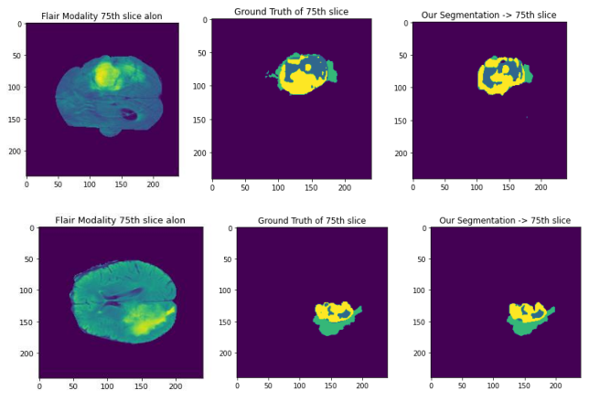
Select a view, open it at full size and compare the original slice, reference annotation and predicted segmentation. Every panel uses a non-cropping contain layout.
FLAIR image, ground truth and model prediction are displayed together for two different views.
Preserves the complete source image and native aspect ratio.
Human-provided class labels serve as the reference annotation.
The model outputs a pixel-wise map for side-by-side review.
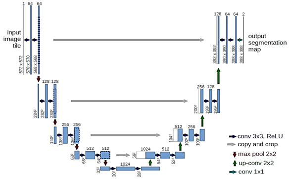
The contracting path learns progressively richer features. The expanding path restores image resolution, while skip connections pass fine spatial information directly to the decoder.
Slice 3D MRI volumes along three anatomical axes.
Encode tissue patterns through convolution and pooling.
Decode features with up-convolution and skip connections.
Fuse axis-specific predictions into a tumor-region map.
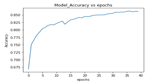
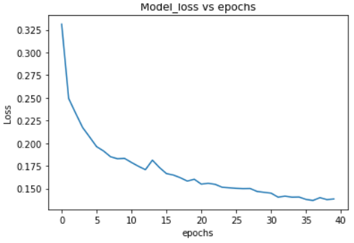
Metrics below reflect the project’s reported improved four-class experiment, presented as research/demo indicators rather than clinical claims.
Presentation disclaimer: NeuroSeg AI is presented as a research and demonstration concept. It is not a certified medical device and must not be used as a substitute for qualified clinical interpretation.
A polished, local-first presentation experience designed to demonstrate how AI can support brain MRI review.