Classification
→
One label
“abnormal”
“abnormal”
Summarizes an entire image into one category, but does not show where the relevant cells are.
 CytoVision AIMicroscopy Intelligence
An interactive presentation of a U-Net based workflow that identifies cell regions in Pap-smear microscopy images, overlays predicted masks, and converts segmentation into measurable morphology.
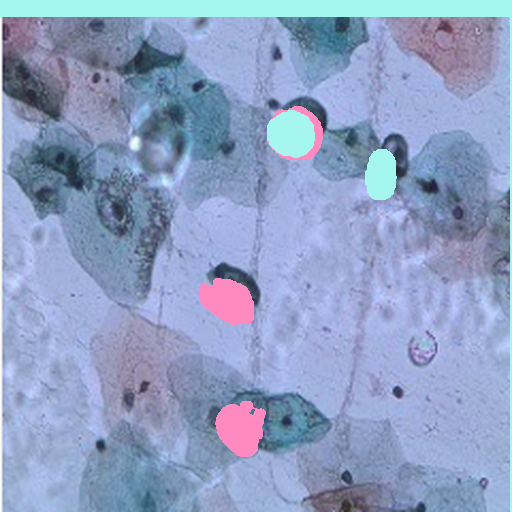
The model produces a spatial map. Every output pixel answers a more useful question: does this location belong to the target cell region?
Summarizes an entire image into one category, but does not show where the relevant cells are.
Produces a binary mask, enabling overlays, area measurements, morphology, quality checks, and human review.
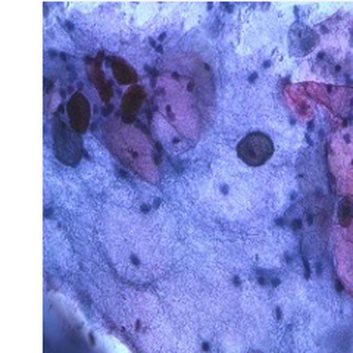
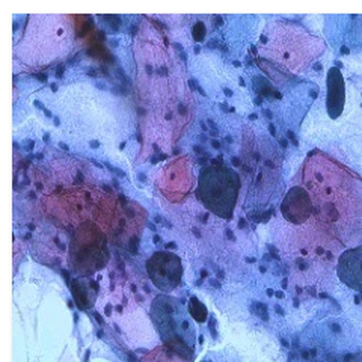
A microscope-mounted camera supplies a color Pap-smear image. The full frame remains available for review while the analysis pathway creates a standardized model input.
This interactive panel demonstrates how a predicted mask can drive practical cell-level measurements. Values are presentation simulations derived from the selected sample, not clinical results.
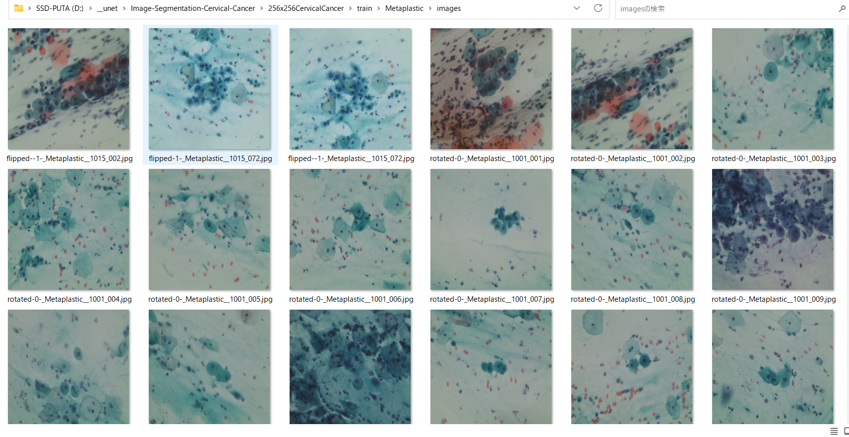
The source microscopy database contains five cervical-cell categories. This experiment prepares image–mask pairs and focuses its demonstrated segmentation training on the Metaplastic category.
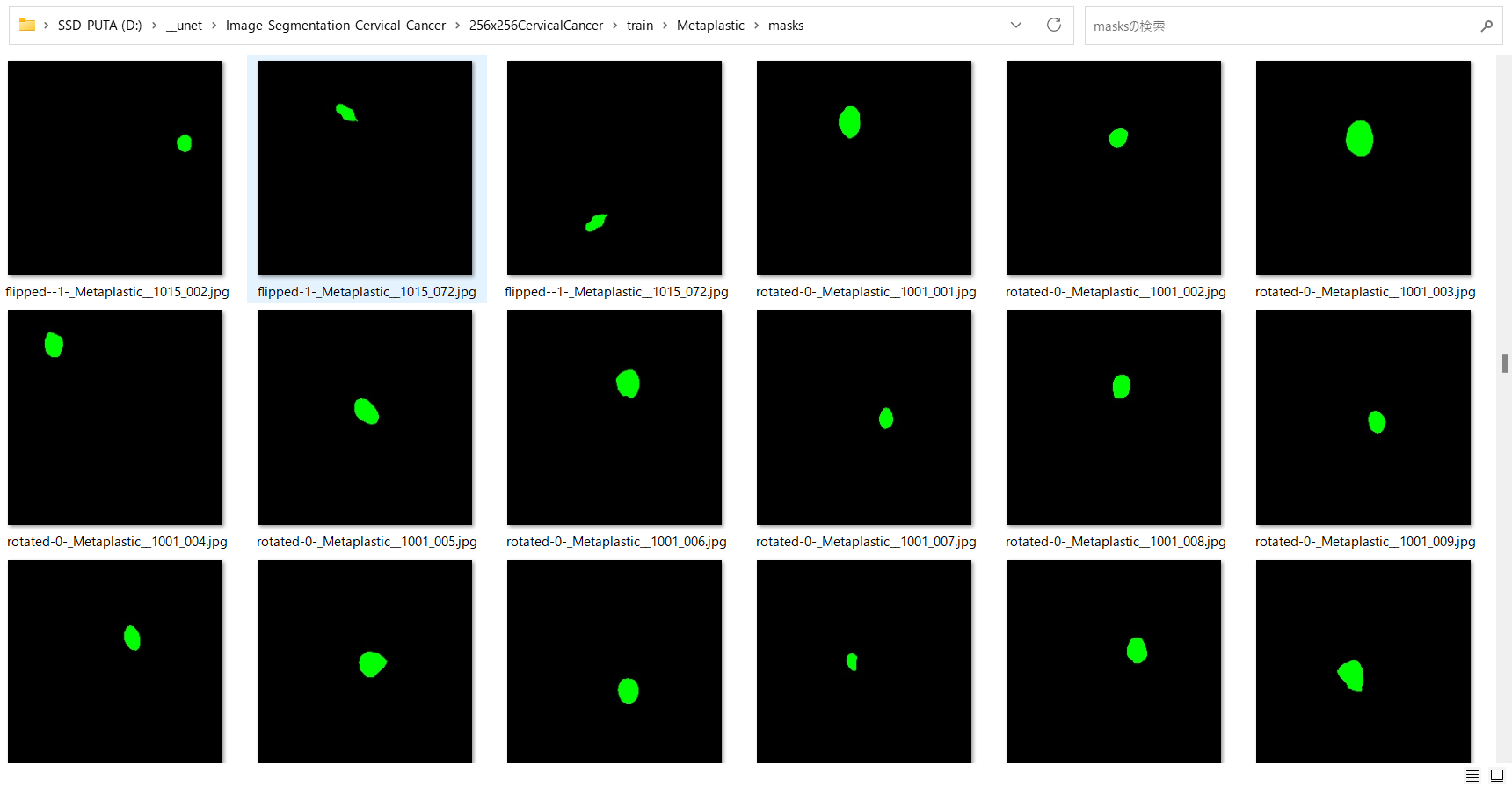
Move the divider to inspect the full image. No scientific media is cropped.
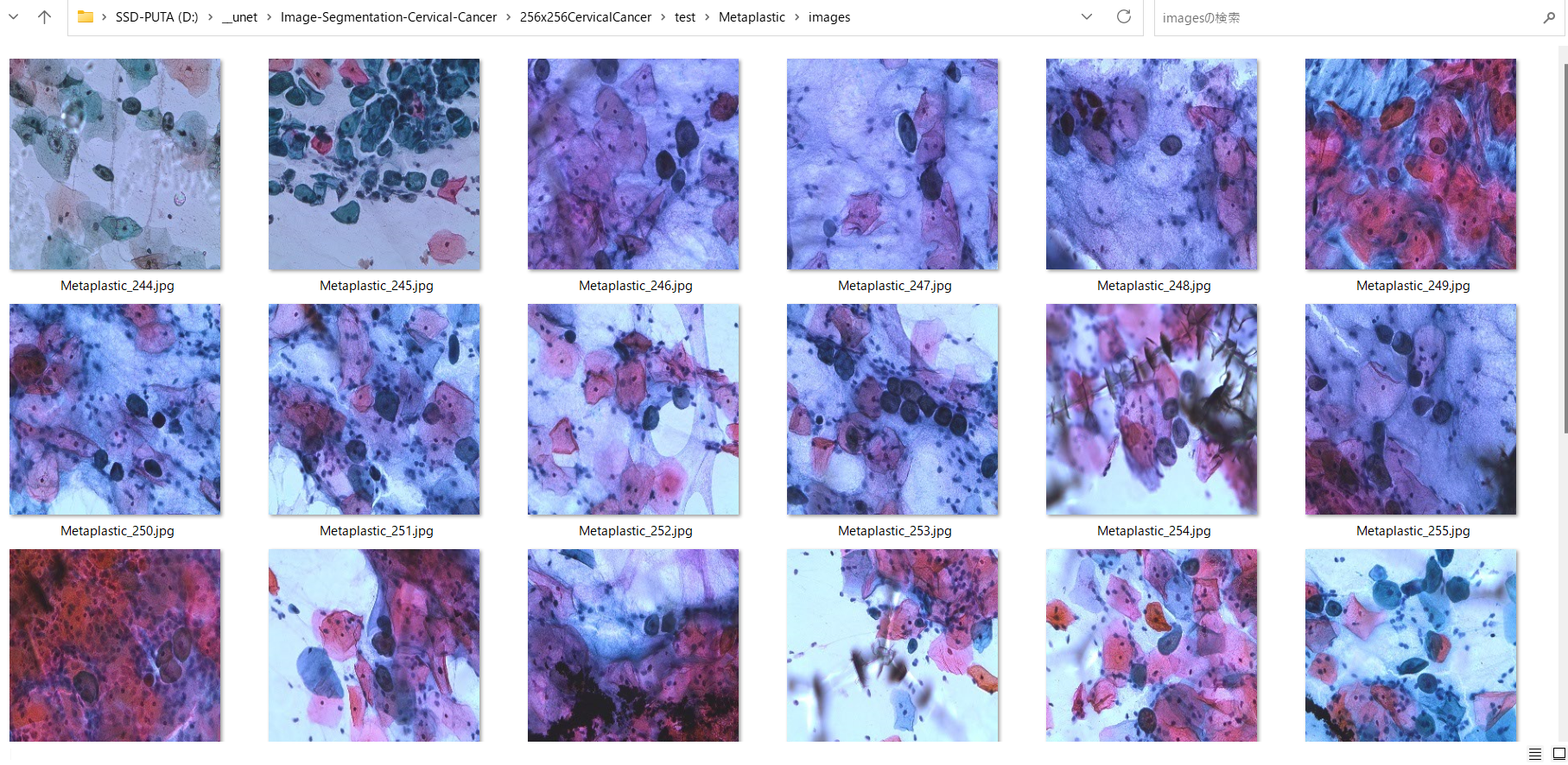
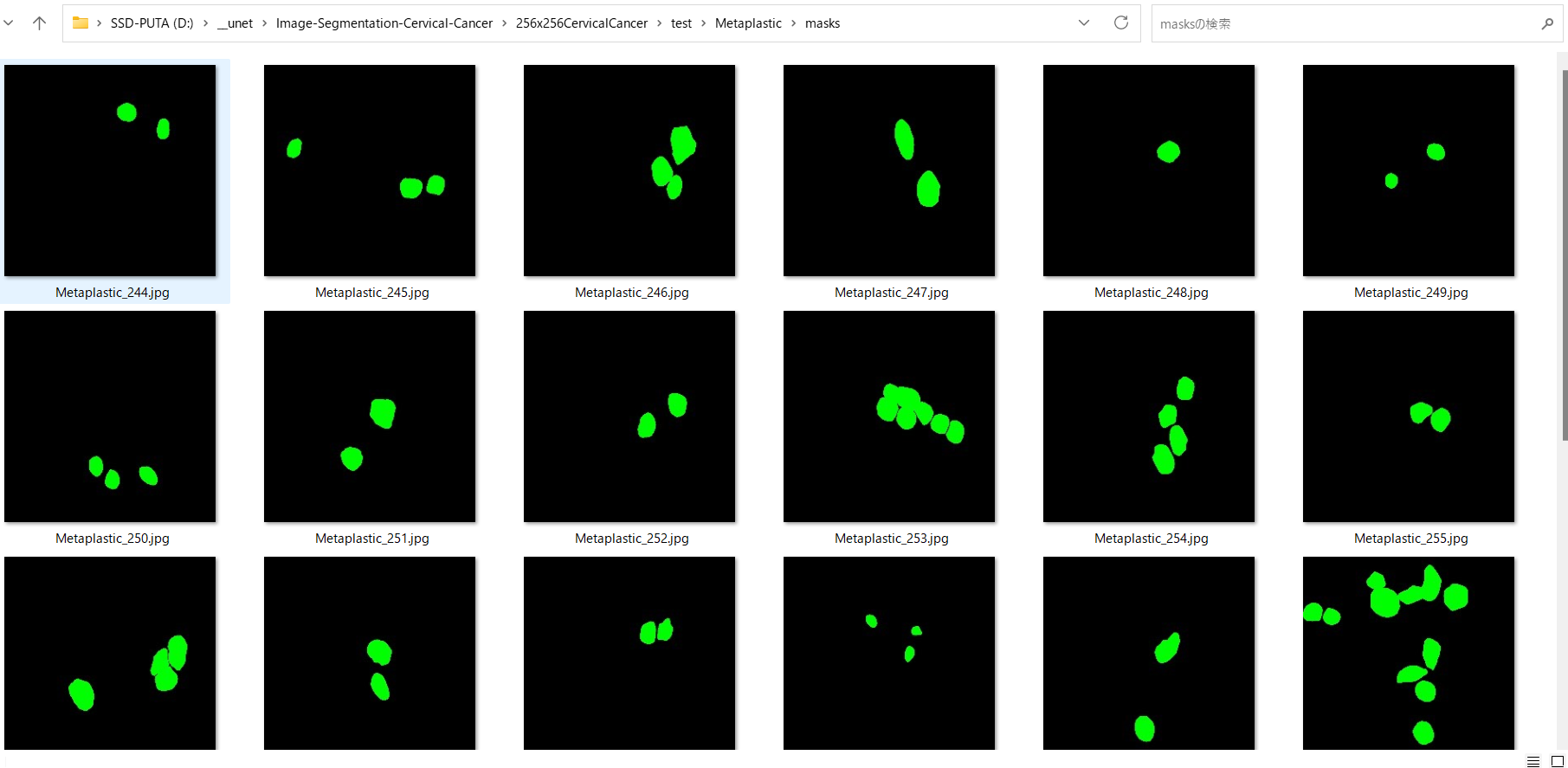
Inspect missed regions, over-segmentation, merged objects and weak edges.
The model assists review; it should not remove expert oversight.
Preserve a traceable record of the input, output and operator response.
The presentation avoids inventing clinical performance. It displays the project’s recorded training artifacts and explains what they do—and do not—establish.
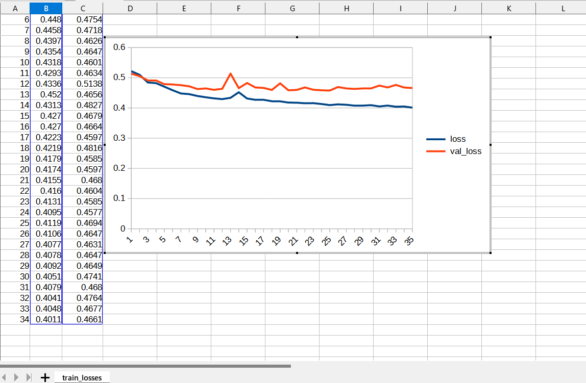
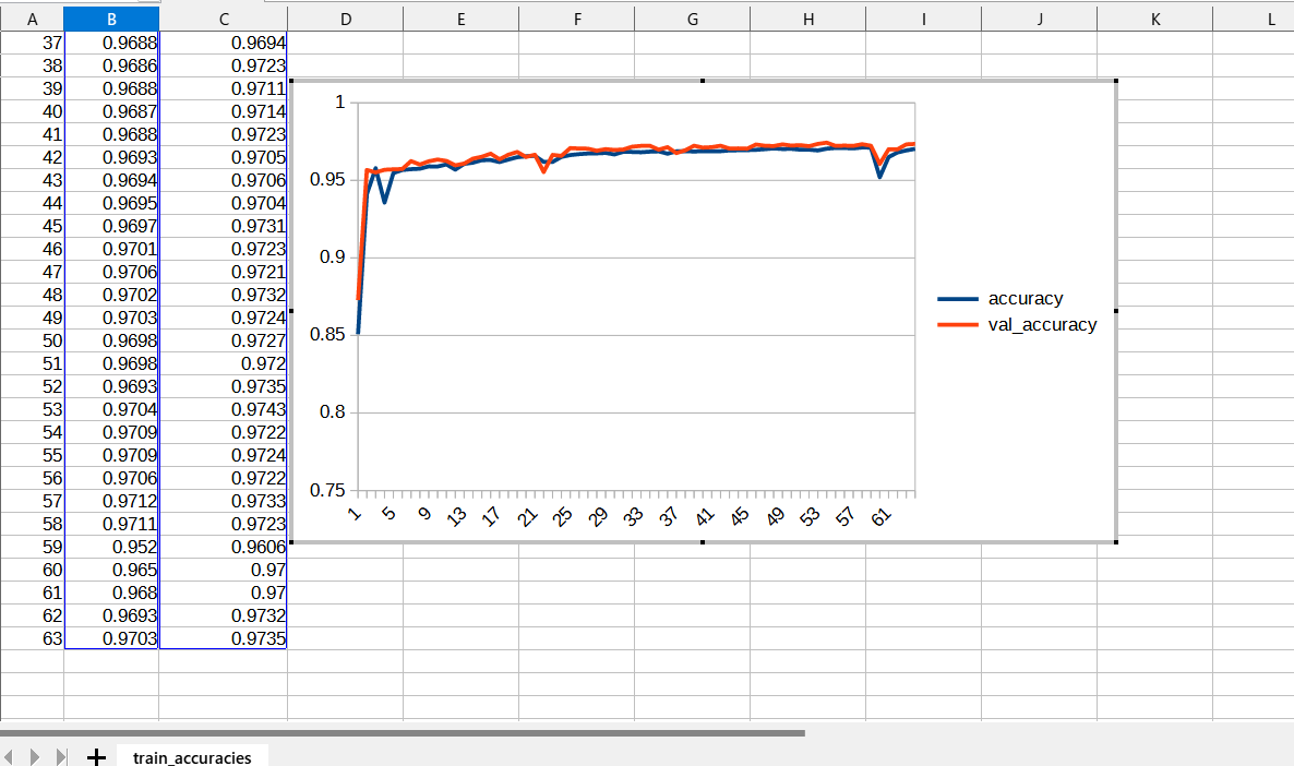
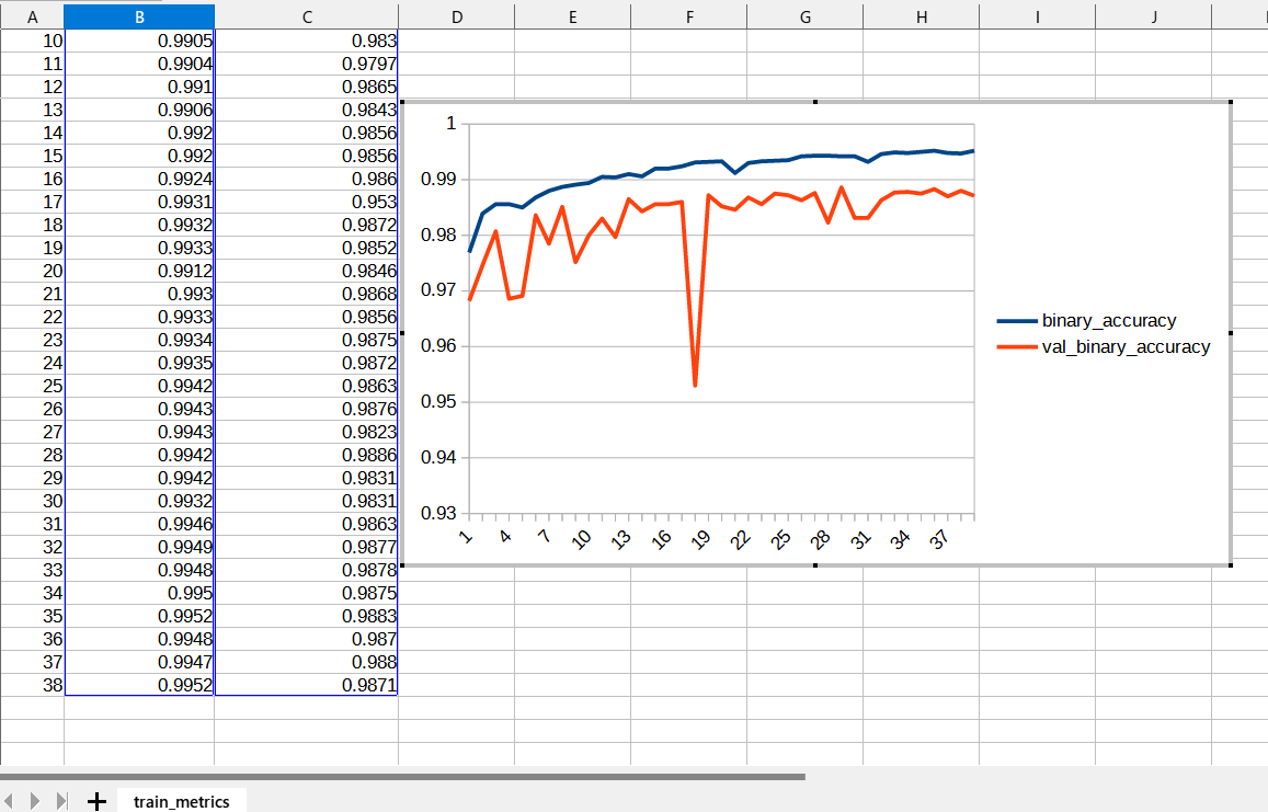
Training curves indicate optimization behavior on the prepared dataset. They do not establish population-level diagnostic accuracy, device performance, or regulatory readiness.
Pair microscope hardware with a visual AI review experience rather than presenting inference as a hidden black box.
Use clear overlays, QC prompts and measurement panels that remain understandable for mixed-skill laboratory teams.
The presentation concept can grow toward multi-cell detection, annotation, batch review, reports and local integrations.
Research presentation for AI-assisted cervical cytology workflows.