01 Non-contrast input
AI-assisted non-contrast CT analysis
AortaGuard AI
See vascular risk signals hidden inside non-contrast CT.
A cascaded 3D intelligence workflow that isolates the aorta, creates a contrast-like representation, estimates lumen structure, and predicts whether the volume is consistent with aortic dissection.
Powered with ❤️ by Solicitous ®
0.938Reported sensitivity
0.847Reported F1 score
3DVolumetric processing
02 AI contrast-like output
AD
MODEL DECISION
Dissection pattern detected
Decision support signal generated from the isolated 3D aortic volume.
01
THE CLINICAL GAP
Non-contrast CT can hide subtle dissection signs.
The aortic lumen and intimal flap are far easier to inspect after contrast enhancement, yet contrast is not suitable for every patient.
02
THE AI STRATEGY
Make the aorta easier for the model to reason about.
Rather than classifying the full scan directly, the system first isolates the aorta and then learns several related vascular tasks together.
03
THE OUTPUT
One input, multiple clinically meaningful signals.
The pipeline produces a contrast-like volume, lumen segmentation cues, and an AD versus non-AD classification.
What is actually happening?
A five-stage cascade, explained without the jargon.
The framework does not ask one black box to make a diagnosis from an entire chest CT. It narrows the problem step by step.
Full NCE-CT
→
3D U-Net
→
Aorta mask
Mask × CT volume
ROI
Only the aortic region moves forward.
This suppresses unrelated lungs, spine, ribs, and soft tissue, giving the next model a focused vascular target.
Generator
Synthetic CE-like volume
True lumenFalse lumenIntimal flap
STRUCTURE
The model learns the anatomy that defines dissection.
Aortic dissection creates an intimal flap that divides blood flow into a true lumen and a false lumen.
CLASSIFIER OUTPUTAortic dissection
Decision informed by the same representation used for synthesis and segmentation.
Stage 01
A pre-trained 3D segmentation network finds the aorta.
The raw non-contrast scan is converted into a clean anatomical mask before diagnosis begins.
Synchronized volumetric viewer
Watch the model transform an entire 3D aortic volume.
Each panel sweeps through the same kind of volumetric data. Switch between a normal case and an aortic-dissection case, pause at any point, or scrub through the scan.
AD CASEIrregular lumen morphology visible across the volume
SYNTHESIZED
Non-contrast CT is the only scan needed at inference time.
Real contrast CT is used as a learning target during development.
Synthetic contrast-like CT is generated to expose vascular structure to the downstream tasks.
Reading the anatomy
What the model is looking for inside the aorta.
The diagnosis becomes more interpretable when the model is trained to represent the anatomical consequences of a dissection.
True lumenNative blood-flow channel
False lumenNew channel created by dissection
Intimal flapTorn inner wall separating the lumens
The aortic wall splits.
Blood enters through a tear in the inner wall and creates a second passage within the vessel wall.
Two lumen patterns emerge.
The normal channel becomes the true lumen; the new dissected channel is the false lumen.
The intimal flap becomes a key cue.
The flap and the shape relationship between both lumens are major structural signals for detection.
Multi-task learning forces anatomical reasoning.
By learning image synthesis, lumen segmentation, and classification together, the shared representation has to capture more than a superficial texture difference.
System architecture
One shared 3D representation, three coordinated objectives.
The generator creates the contrast-like volume and lumen map. A classifier predicts AD versus non-AD, while a discriminator pushes the generated output toward realistic paired contrast data.
INPUTNCE-CT volumeNon-contrast 3D scan
STAGE 13D aorta maskPre-trained anatomical locator
STAGE 2Multi-task generatorShared 3D feature learning
OUTPUT ACE-like volumeVascular image synthesis
OUTPUT BLumen mapTrue / false lumen segmentation
OUTPUT CAD decisionPatient-level classification
Discriminator
Checks whether the generated contrast-like volume resembles the real paired distribution.
Shared learning reduces shortcut behaviour.
The classifier, image-synthesis task, and lumen-segmentation task all depend on the same hidden representation. Useful features therefore need to describe the aortic structure, not merely one isolated visual pattern.
ORIGINAL SCIENTIFIC OVERVIEW
Complete pipeline visual abstract
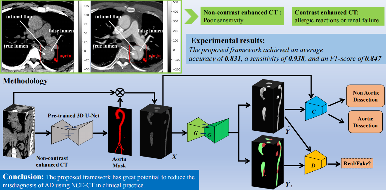
Reported research results
Designed to prioritize sensitivity for a dangerous, easily missed condition.
These values come from the published experimental evaluation and should be presented as research performance, not as a regulatory or clinical-deployment claim.
Average accuracy
Overall proportion of AD and non-AD cases classified correctly.
Sensitivity
Ability to identify patients with aortic dissection in the reported evaluation.
F1 score
Balance between positive-case precision and sensitivity.
The visual is conceptual; the exact metric reported by the study was 0.938.
Why sensitivity matters here
Aortic dissection is time-critical. In a screening or decision-support context, missing a true case can be especially harmful. The study therefore highlights sensitivity alongside accuracy and F1 score.
!
Research-use notice: The source implementation is a research framework. Clinical use would require independent validation, safety engineering, regulatory review, and integration into an approved workflow.
Presentation workflow
How this can be demonstrated as an instrument-connected AI experience.
A polished product layer can turn the research pipeline into a clear, operator-friendly local demonstration.
Import CT volume
Load a paired or non-contrast NIfTI volume into the workstation.
Localize the aorta
Run the 3D anatomical segmentation stage to isolate the vascular region.
Generate vascular cues
Create the contrast-like representation and lumen segmentation outputs.
Review the decision
Inspect synchronized volumes, structural masks, and the patient-level prediction.
Export a report
Generate a presentation-ready summary with images, metrics, and model notice.
Volumetric segmentation, conditional synthesis, adversarial quality control, and classification.
The repository describes a standardized size of 256 × 256 × 512 voxels for paired datasets.
Training and testing scripts use a 3D pix2pix-inspired paired-data workflow.
Best presented as a research and instrument-integration prototype, not a standalone diagnosis.

AortaGuard AI
From a difficult non-contrast scan to a structured vascular explanation.
Built to demonstrate how generative AI, segmentation, and classification can work together inside one focused medical-imaging experience.
Powered with ❤️ by Solicitous ®